
Exercise therapy in diseases of the peripheral nervous system. Exercise therapy in diseases of the central nervous system. Depending on the location, there are
Nervous system controls the activities of various organs and systems that make up the whole organism, communicates with the external environment, and also coordinates the processes occurring in the body depending on the state of the external and internal environment. It coordinates blood circulation, lymph flow, metabolic processes, which, in turn, affect the state and activity nervous system.
The human nervous system is conditionally divided into central and peripheral (Fig. 121). In all organs and tissues, nerve fibers form sensory and motor nerve endings. The first, or receptors, provide the perception of irritation from the external or internal environment and convert the energy of stimuli (mechanical, chemical, thermal, light, sound, etc.) in the process of excitation, which is transmitted to the central nervous system. Motor nerve endings transmit excitation from the nerve fiber to the innervated organ.
Rice. 121. Central and peripheral nervous system.
A: 1 - phrenic nerve;2 - brachial plexus;3 - intercostal nerves;4 - axillary nerve;5 - musculocutaneous nerve;6 - radial nerve;7 - median nerve;8 - ulnar nerve;9 - lumbar plexus;10 - sacral plexus;11 - pudendal and coccygeal plexus;12 - sciatic nerve;13 - peroneal nerve;14 - tibial nerve;15 - brain;16 - external cutaneous nerve of the thigh;17 - lateral dorsal cutaneous nerve;18 - tibial nerve.
B - segments of the spinal cord.
B - spinal cord:1 - white matter;2 - gray
substance;3 - spinal canal;4 - front horn;5 -
rear horn;6 - front roots;7 - back roots;8 -
spinal node;9 - spinal nerve.
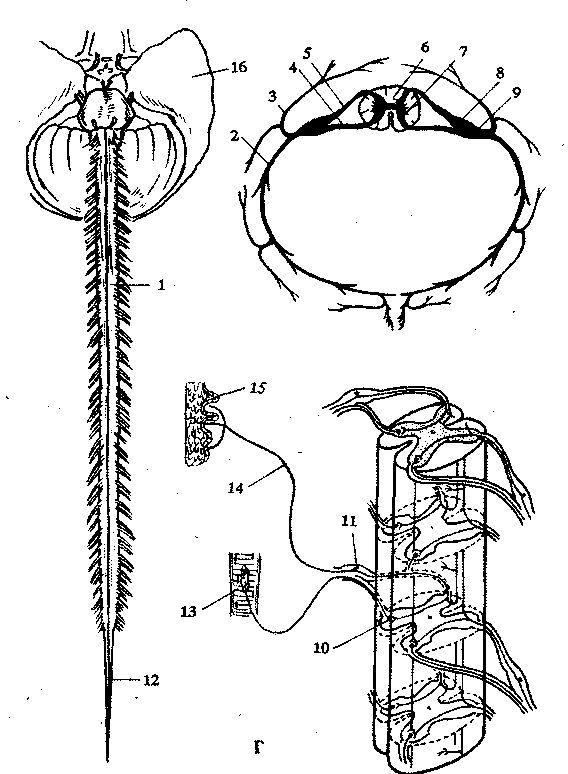
G: 1 - spinal cord;2 - anterior branch of the spinal nerve;3 - posterior branch of the spinal nerve;4 - anterior root of the spinal nerve;5 - posterior root of the spinal nerve;6 - rear horn;7 - front horn;8 - spinal node;9 - spinal nerve;10 - motor nerve cell;11 - spinal node;12 - terminal thread;13 - muscle fibers;14 - sensitive nerve;15 - the end of the sensory nerve,16 - brain
It is known that higher motor centers are located in the so-called motor zone of the cerebral cortex - in the anterior central gyrus and adjacent areas. Nerve fibers from the indicated region of the cerebral cortex pass through the inner capsule, the subcortical regions and at the border of the brain and spinal cord make an incomplete decussation with the transition of most of them to the opposite side. Therefore, in diseases of the brain, motor disorders are observed on the opposite side: when the right hemisphere of the brain is damaged, the left half of the body is paralyzed, and vice versa. Further, the nerve fibers descend as part of the bundles of the spinal cord, approaching the motor cells, motoneurons of the anterior horns of the spinal cord. Motor neurons that regulate the movements of the upper limbs lie in the cervical thickening of the spinal cord (level V-VIII of the cervical and I-II thoracic segments), and lower extremities- in the lumbar level I-V lumbar and I-II sacral segments). The fibers coming from the nerve cells of the nuclei of the base nodes - the subcortical motor centers of the brain, from the reticular formation of the brain stem and cerebellum are also sent to the same spinal motor neurons. Thanks to this, the regulation of coordination of movements is ensured, involuntary (automated) movements are carried out and voluntary movements are prepared. The fibers of the motor cells of the anterior horns of the spinal cord, which are part of the nerve plexuses and peripheral nerves, end in the muscles (Fig. 122).

Rice. 122. Dermatome boundaries and segmental innervation(A, B), muscles
human(B), transverse section of the spinal cord(G).
A: C 1-8 - cervical;T 1-12 - chest;L1-5 - lumbar;S 1-5 - sacral.
B: 1 - cervical knot;2 - median cervical node;3 -
lower cervical node;4 - border sympathetic trunk;
5 - cerebral cone;6 - terminal (terminal) thread
meninges;7 - lower sacral node
sympathetic trunk.
B (front view):1 - frontal muscle;2 - chewing
muscle; 3 - sternocleidomastoid muscle;4 -
pectoralis major;5 - the latissimus dorsi muscle;6 -
serratus anterior;7 - white line;8 - seed
cord;9 - flexor thumb brushes;10 -
quadriceps femoris;11 - long fibula
muscle;12 - anterior tibialis muscle;13 - long
extensor of fingers;14 - short muscles of the rear of the foot;15 -
facial muscles;16 - subcutaneous muscle of the neck;
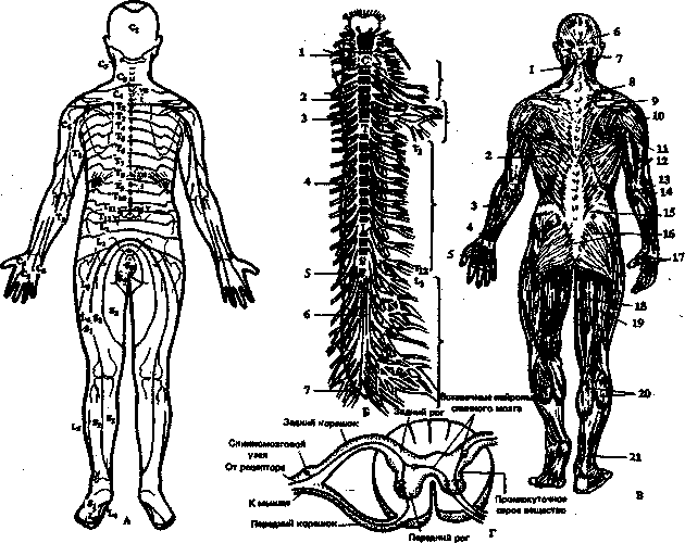
17 - collarbone;18 - deltoid muscle;19 - sternum;20 - biceps muscle of the shoulder;21 - rectus abdominis;22 - muscles of the forearm;23 - umbilical ring;24 - worm-like muscles;25 - wide fascia of the thigh;26 - adductor muscle of the thigh;27 - tailor muscle;28 - extensor tendon retainer;29 - long extensor of the fingers;30 - external oblique muscle of the abdomen.
B (back view):1 - belt muscle of the head;2 - the latissimus dorsi muscle; 3 - ulnar extensor of the wrist;4 - extensor of the fingers;5 - muscles of the rear of the hand;6 - tendon helmet;7 - external occipital protrusion;8 - trapezius muscle;9 - spine of the scapula;10 - deltoid muscle;11 - rhomboid muscle;12 - triceps muscle of the shoulder;13 - medial epicondyle;14 - long radial extensor of the wrist;15 - chest-lumbar fascia;16 - gluteal muscles;17 - muscles of the palmar surface of the hand;18 - semimembranous muscle;19 - biceps;20 - calf muscle;21 - Achilles (heel) tendon
Any motor act occurs when an impulse is transmitted along the nerve fibers from the cerebral cortex to the anterior horns of the spinal cord and further to the muscles (see Fig. 220). In diseases (injuries of the spinal cord) of the nervous system, the conduction of nerve impulses becomes difficult, and a violation of the motor function of the muscles occurs. Complete loss of muscle function is called paralysis (plegia), and the partial paresis.
According to the prevalence of paralysis, there are: monoplegia(lack of movement in one limb - arm or leg), hemiplegia(damage to the upper and lower limbs of one side of the body: right-sided or left-sided hemiplegia), paraplegia(impaired movement in both lower limbs is called lower paraplegia, in the upper - upper paraplegia) and tetraplegia (paralysis of all four limbs). When peripheral nerves are damaged, paresis in the zone of their innervation, called the corresponding nerve (for example, paresis of the facial nerve, paresis of the radial nerve, etc.) (Fig. 123).
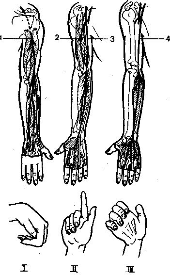
Rice. 123. Nerves of the upper limb;1 - radial nerve;2 - skin-
muscular nerve;3 - median nerve;4 - ulnar nerve.I - brush with damage to the radial nerve.II - brush with damage to the median nerve.III - hand with damage to the ulnar nerve
Depending on the localization of the lesion of the nervous system, peripheral or central paralysis (paresis) occurs.
With the defeat of the motor cells of the anterior horns of the spinal cord, as well as the fibers of these cells, which are part of the nerve plexuses and peripheral nerves, a picture of peripheral (flaccid), paralysis develops, which is characterized by a predominance of symptoms of neuromuscular prolapse: limitation or absence of voluntary movements, a decrease in muscle strength, decreased muscle tone (hypotension), tendon, periosteal and skin reflexes (hyporeflexia) or their complete absence. Often there is also a decrease in sensitivity and trophic disorders, in particular muscle atrophy.
To correctly determine the severity of paresis, and in cases of mild paresis - sometimes to identify it, it is important to quantify the state of individual motor functions: muscle tone and strength, and the volume of active movements. The available methods make it possible to compare and effectively control the results of rehabilitation treatment in a polyclinic and a hospital.
To study muscle tone, a tonometer is used, muscle strength is measured with a hand dynamometer, and the volume of active movements is measured with a goniometer (in degrees).
In case of violation of the cortical-subcortical connections with the reticular formation of the brain stem or damage to the descending motor pathways in the spinal cord and, as a result, the function of the spinal motor neurons is activated as a result of a disease or brain injury, a syndrome of central spastic paralysis occurs. It, in contrast to peripheral and central "flaccid" paralysis, is characterized by an increase in tendon and periosteal reflexes (hyperflexia), the appearance of pathological reflexes, the occurrence of the same movements when trying to voluntarily act on a healthy or paralyzed limb (for example, abduction of the shoulder outward when bending the forearm of the paretic hands or clenching a paralyzed hand into a fist with a similar voluntary movement of a healthy hand).
One of the most important symptoms of central paralysis is a pronounced increase in muscle tone (muscle hypertension), which is why such paralysis is often called spastic. For most patients with central paralysis due to brain disease or injury, the Wernicke-Mann posture is characteristic: the shoulder is brought (pressed) to the body, the hand and forearm are bent, the hand is turned palm down, and the leg is extended at the hip and knee joints and bent at the foot. This reflects a predominant increase in the tone of the flexor and pronator muscles in the upper limb and extensor muscles in the lower.
With injuries and diseases of the nervous system, disorders occur that sharply reduce the efficiency of patients, often lead to the development of secondary paralytic deformities and contractures that adversely affect the musculoskeletal function. Common to all injuries and diseases of the nervous system are limitation of the range of motion, decreased muscle tone, vegetotrophic disorders, etc.
A deep understanding of the mechanisms of the pathology of the nervous system is the key to the success of rehabilitation measures. So, with discogenic sciatica, nerve fibers are infringed, causing pain, with a stroke, certain areas of motor nerve cells cease to function, so adaptation mechanisms play an important role.
In rehabilitation, compensatory-adaptive reactions of the body are important, which are characterized by the following common features: normal physiological functions of organs and tissues (their functions); adaptation of the organism to the environment, provided by the restructuring of vital activity due to the strengthening of some and the simultaneous weakening of other of its functions; they develop on a single, stereotyped material basis in the form of continuous variation in the intensity of renewal and hyperplasia of the cellular composition of tissues and intracellular structures; compensatory-adaptive reactions are often accompanied by the appearance of peculiar tissue (morphological) changes.
The development of regenerative processes in the nervous tissue occurs under the influence of preserved functions, that is, the nervous tissue is being restructured, the number of processes of nerve cells and their branches on the periphery changes; there is also a restructuring of synaptic connections and compensation after the death of part of the nerve cells.
The process of restoration of the nervous system occurs in nerve cells, nerve fibers and structural elements of tissues due to (or due to) restoration of membrane permeability and excitability, normalization of intracellular redox processes and activation of enzyme systems, which leads to the restoration of conductivity along nerve fibers and synapses.
The rehabilitation regimen should be adequate to the severity of the disease, which is assessed by the degree of impairment of adaptive activity. The level of damage to the central nervous system and peripheral nervous system is taken into account. Important factors are the ability to move independently, take care of oneself (perform housework, eat alone, etc.) and family, communicate with others, assess the adequacy of behavior, the ability to control physiological functions, as well as the effectiveness of training.
A comprehensive rehabilitation system includes the use of exercise therapy, hydrokinesitherapy, different kinds massage, occupational therapy, physiotherapy, spa treatment, etc. In each individual case, the combination and sequence of the use of certain means of rehabilitation is determined.
In case of severe diseases (injuries) of the nervous system, rehabilitation is aimed at improving general condition patients, raising their emotional tone and forming their correct attitude to the prescribed treatment and the environment: psychotherapy, symptomatic drug therapy, occupational therapy, music therapy, massage in combination with therapeutic exercises, etc.
Exercise therapy in neurology has a number of rules, the observance of which makes this method the most effective: early use of exercise therapy; the use of its means and techniques to restore temporarily impaired functions or to maximize compensation for those lost; selection of special exercises in combination with general developmental, general strengthening exercises and massage; strict individuality of exercise therapy, depending on the diagnosis, age and gender of the patient; active and steady expansion of the motor mode from the lying position to the transition to the sitting position, standing, etc.
Special exercises can be conditionally divided into the following groups:
exercises that increase joint range of motion and muscle strength;
exercises aimed at restoring and improving coordination of movements;
antispastic and antirigid exercises;
ideomotor exercises (sending a mental impulse to a trained muscle group);
a group of exercises aimed at restoring or forming motor skills (standing, walking, manipulations with simple but important household objects: clothes, dishes, etc.);
passive exercises and exercises for stretching connective tissue formations, treatment with position, etc.
All of the above groups of exercises are combined in various combinations and depend on the nature and extent of the motor defect, the stage of rehabilitation, the age and gender of the patient.
Rehabilitation of neurological patients requires long-term training of compensatory mechanisms (walking on crutches, self-care, etc.) to ensure sufficient compensation for lost or impaired functions. However, at a certain stage (stages), the recovery process slows down, that is, stabilization occurs. The success of rehabilitation is different for a particular pathology. So, with osteochondrosis of the spine or lumbosacral sciatica, it is higher than with multiple sclerosis or vascular disease.
Rehabilitation largely depends on the patient himself, on how diligently he performs the program prescribed by the rehabilitation doctor or exercise therapy methodologist, helps to correct it depending on his functional capabilities, and, finally, whether he continues recovery exercises after the rehabilitation period is over.
Brain injury (concussion)
All brain injuries are characterized by an increase in intracranial pressure, a violation of hemo- and liquor circulation, followed by a violation of cortical-subcortical neurodynamics with macro- and microscopic changes in the cellular elements of the brain. A concussion of the brain leads to headaches, dizziness, functional and persistent autonomic disorders.
In case of violations of motor functions for the prevention of contractures, exercise therapy is prescribed (passive, then passive-active movements, positional treatment, muscle stretching exercises, etc.), massage of the back and paralyzed limbs (first the legs are massaged, then the arms, starting from the proximal sections), and also affect the biologically active points (BAP) of the limbs.
With mild and moderate concussion, massage should be carried out from the second or third day after the injury in the patient's sitting position. First, the back of the head, neck, shoulder girdle are massaged, then the back to the lower corners of the shoulder blades, using stroking, rubbing, shallow kneading and light vibration. Finish the procedure by stroking from the scalp to the muscles of the shoulder girdle. The duration of the massage is 5-10 minutes. Course 8-10 procedures.
In the first 3-5 days, with mild to moderate concussion, cryomassage of the occipital region and muscles of the shoulder girdle is also used. The duration of the massage is 3-5 minutes. Course 8-10 procedures.
Injuries of the spine and spinal cord
Sometimes a spinal injury occurs in a position of hyperlordosis, and then a rupture of an intact intervertebral disc can occur.
The cervical spine is especially often injured when jumping into a shallow body of water, when, after hitting the head against the bottom, a traumatic prolapse of an intact intervertebral disc occurs, causing tritraplegia. Degenerative changes inevitably lead to herniation of the intervertebral discs, which in itself is not a cause for complaints, but due to trauma, a radicular syndrome occurs.
When the spinal cord is damaged, flaccid paralysis occurs, which is characterized by muscle atrophy, the impossibility of voluntary movements, the absence of reflexes, etc. Each muscle is innervated from several segments of the spinal cord (see Fig. 96), therefore, with damage or diseases, there may be not only paralysis, but also muscle paresis of varying severity, depending on the prevalence of lesions in the anterior horns of the gray matter of the spinal cord.
The clinical course of the disease depends on the degree of damage to the spinal cord and its roots (see Fig. 122). So, with injuries of the upper cervical of the spine, spastic tetraparesis of the extremities occurs. With lower cervical and upper thoracic localization (C 6 -T 4), flaccid paresis of the arms and spastic paresis of the legs occurs, with thoracic localization - paresis of the legs. With the defeat of the lower thoracic and lumbar segments of the spine, flaccid paralysis of the legs develops. The cause of flaccid paralysis can also be damage to the spinal cord with closed fractures of the spine and its injuries.
Prevention of the development of joint contractures by means of massage, exercise therapy, stretching exercises, physio- and hydrotherapy, hydrokinesitherapy is the main task for paralysis of any origin. In water, the possibility of active movements is facilitated and the fatigue of weakened muscles is reduced. Electrical stimulation of paralyzed muscles is carried out with needle electrodes with a preliminary introduction of ATP. In addition, positional treatment is included using staged plaster splints (bandages), teips, sandbags, etc., as well as staged redressing and other methods.
Timely use of the necessary rehabilitation means can completely prevent the development of contractures and other deformities.
Traumatic encephalopathy is a complex of morphological, neurological and mental disorders that occur in the late and long-term periods after a traumatic brain injury. Characterized by asthenic and various vegetative-vascular disorders, memory impairment by the type of retrograde amnesia, headaches, fatigue, irritability, sleep disturbance, heat intolerance, stuffiness, etc.
The recurrence of seizures indicates the development of traumatic epilepsy. In severe cases, traumatic dementia occurs with severe memory impairment, a decrease in the level of personality, etc.
In addition to dehydration therapy, complex treatment includes the use of anticonvulsants, tranquilizers, nootropics, etc. Massage, LH, walking, skiing help to improve the patient's well-being and prevent the onset of decompensation.
The massage technique includes massaging the collar area, back (to the lower corners of the shoulder blades), legs, as well as the effect on the BAP by the inhibitory or stimulating method, depending on the prevalence of one or another symptom. The duration of the massage is 10-15 minutes. Course 10-15 procedures. 2-3 courses per year. With a headache, cryomassage No. 5 is indicated.
Patients are not allowed to visit the bath (sauna), sunbathe, take hyperthermic baths!
Vascular epilepsy
The occurrence of epileptic seizures in dysciculatory encephalopathy is associated with the formation of cicatricial and cystic changes in the brain tissue and regional cerebral hypoxia.
The system of rehabilitation of patients includes exercise therapy: general developmental exercises, breathing, coordination. Exercises with straining, with weights, as well as with prolonged head tilts are excluded. Therapeutic exercises are performed at a slow pace, without sudden movements. Swimming, cycling, visiting the sauna (bath) are also excluded.
Physiotherapy includes electrosleep, drug electrophoresis No. 10, oxygen therapy. A general massage is performed, with the exception of percussion techniques. Occupational therapy is carried out on stands, box gluing, bookbinding, etc.
Osteocondritis of the spine
Degenerative changes in the intervertebral discs occur as a result of the physiological neuroendocrine aging process and due to wear and tear under the influence of one-time injuries or repeated microtraumas. Most often, osteochondrosis occurs in athletes, hammerers, typists, weavers, drivers, machine operators, etc.
Speedy recovery of function spinal column general massage, cryomassage, vibration massage, LG (Fig. 124), hydrocolonotherapy help. They cause deep hyperemia, improve blood and lymph flow, have an analgesic and resolving effect.
Massage technique. First, a preliminary back massage is performed using stroking techniques, shallow kneading of the muscles of the entire back. Then they proceed to massage the spinal column, using rubbing with the phalanges of four fingers, the base of the palm, kneading with the phalanges of the first fingers, forceps, ordinary and double ring kneading of the broad muscles of the back. Particularly carefully grind, knead BAP. Rubbing and kneading techniques should be alternated with stroking with both hands. In conclusion, active-passive movements are carried out, breathing exercises with an emphasis on exhalation and compression of the chest 6-8 times. The duration of the massage is 10-15 minutes. Course 15-20 procedures.

Rice. 124. Approximate complex of LH in osteochondrosis of the spine
Discogenic radiculitis
The disease often affects the intervertebral discs of the lower part of the spinal column. This is explained by the fact that the lumbar region has greater mobility and is subjected to the most intense static-dynamic loads on the muscular-ligamentous apparatus. Pain occurs when the spinal nerve roots are compressed by a disc herniation. The pain syndrome is characterized by acute development. Pain can occur in the morning, after heavy physical exertion, and in some cases is accompanied by muscle spasm. There is some limitation of movements in the lumbar spine, lumbar discomfort.
Conservative treatment is shown. Traction is carried out on the shield with a preliminary massage or heating with a solar lamp or manual therapy. After the disappearance of pain - LH in the prone position, on all fours, in the knee-elbow position. The pace is slow to avoid pain. Exercises with inclinations in a standing position are excluded.
Massage objectives: to provide analgesic and anti-inflammatory effects, to promote the speedy recovery of spinal function.
Massage technique. First, stroking, light vibration is performed in order to relieve tension in muscle tone, then longitudinal and transverse kneading of the broad muscles of the back, rubbing with fingertips along the spinal column. Tapping, chopping should not be used to avoid muscle spasm and increased pain. After the procedure, traction is carried out on a shield or in water. The duration of the massage is 8-10 minutes. Course 15-20 procedures.
lumbosacral pain spinal injuries occur, as a rule, immediately after a fall, blow, etc. In mild cases, transient lumbodynia develops with pain in the lumbar region. Acute pain can result from excessive flexion in the lumbosacral region.
LH is performed in the supine position. Includes exercises to stretch the sciatic nerve. Raising the legs up 5-8 times; "bicycle" 15-30 s; twists bent at the knees and hip joints legs left and right 8-12 times; raise the pelvis, pause for a count of 5-8, then return to the starting position. The last exercise is diaphragmatic breathing.
Massage objectives: to provide analgesic and anti-inflammatory effects, improve blood and lymph flow in the damaged area.
Massage technique. The initial position of the patient is lying on his stomach, a roller is placed under the ankle joints. Planar and embracing stroking is applied with the palms of both hands. Kneading is performed with both hands both longitudinally and transversely, while massage movements are performed in ascending and descending directions. In addition, planar stroking is used with the first fingers of both hands in the upward direction, rubbing and kneading with the fingertips, the base of the palm along the spinal column. All massage techniques should be alternated with stroking. Do not use chopping, tapping and intensive kneading. In the early days, the massage should be gentle. The duration of the massage is 8-10 minutes. Course 15-20 procedures.
Lumbago (lumbago) is perhaps the most common manifestation of pain in the lumbar region. Attack-like developing acute piercing pains are localized in the muscles of the lower back and lumbo-dorsal fascia. The disease often occurs in people engaged in physical labor, in athletes, etc., with the combined effect of tension in the lumbar muscles and hypothermia. Chronic infections also play an important role. Pain usually lasts for several days, sometimes 2-3 weeks. Pathophysiologically, with lumbago, there is a tear of the muscle bundles and tendons, hemorrhages in the muscles, and the subsequent phenomena of fibromyositis.
LH (general developmental exercises, stretching exercises and breathing exercises) are performed in the prone position and knee-elbow. The pace is slow. Traction on the shield and cupping massage are shown.
Massage technique. First, a preliminary massage of all the muscles of the back is carried out, then stroking, rubbing and shallow kneading of the muscles of the lumbar region. Professor S.A. Flerov recommends massaging the lower hypogastric sympathetic plexus in the lower abdomen, at the site of bufurcation of the abdominal aorta. Observations show that massage according to the method of S.A. Flerova relieves pain. In the acute period, cryomassage No. 3 is indicated.
sciatica
According to most authors, the disease is caused mainly by congenital or acquired changes in the spinal column and its ligamentous apparatus. Significant and prolonged physical stress, trauma, unfavorable microclimatic conditions, and infections contribute to the development of the disease.
The pain of sciatica can be sharp or dull. It is localized in the lumbosacral region, usually on one side, radiates to the buttock, back of the thigh, outer surface of the lower leg, sometimes combined with numbness, paresthesia. Hyperesthesia is often found
Exercise therapy for diseases, injuries and injuries of the musculoskeletal system and nervous system
Lecture 3exercise therapy for diseases
injuries and injuries
musculoskeletal
apparatus and nervous system
1. Exercise therapy for diseases of the musculoskeletal system
2. Exercise therapy for musculoskeletal injuries
3. Exercise therapy for diseases and injuries of the spine
4. Exercise therapy for diseases and injuries of the nervous system
Question 1. Exercise therapy for diseases of the musculoskeletal system
Tasks of exercise therapy:
normalization of the tone of the central nervous system;activation of metabolism.
activation of blood and lymph circulation in the joint;
restoring or improving joint mobility
prevention of further dysfunctions and
muscle atrophy;
restoration of adaptation to domestic and labor
processes.
Arthritis
are diseases that areis the inflammatory process,
located in the synovium
joint sheath, articular cartilage and
periarticular tissues
Tasks of exercise therapy:
General +increase in range of motion up to
normal;
strengthening muscles in the affected area -
especially extensors;
Exercise therapy technique
1) Therapeutic massage, physiotherapy procedures (UVI,ozokerite, paraffin and mud applications)
2) Therapeutic gymnastics:
I.p .: for the upper limbs - lying and sitting, for the lower - lying
passive movements for affected joints (starting with
gentle swings with a small amplitude)
relaxation of muscles in the area of the diseased joint (relaxation
tense flexor muscles of the diseased limb contributes to
performing active movements with a healthy limb)
exercises in water (in the pool, bath) at a temperature of 28-29 ° C:
active movement,
with shells (ladder for developing movements in the joints
brushes, clubs, dumbbells weighing 0.5 kg), on the gymnastic wall;
simulators.
The pace of the exercises is slow or medium;
Number of repetitions - 12-14 times (14-16 times)
Duration of the lesson - 35-40 minutes (40-45 minutes)
Arthrosis
are diseases that are based onmetabolic-dystrophic process,
characterized by cartilage atrophy,
loss of bone tissue (osteoporosis),
neoplasm of bone tissue
calcium salts in periarticular tissues, ligaments,
joint capsule.
Tasks of exercise therapy:
General +pain reduction;
relaxation of the abdominal muscles and
elimination of contracture;
an increase in the joint space;
reduction of the phenomena of aseptic synovitis
(inflammation of the synovial membrane);
strengthening of the periarticular muscles and increase
their endurance;
Exercise therapy technique
1) Exercises that strengthen the muscles of the back and abdomen.2) Special exercises
i.p. - lying on your back:
active dynamic exercises for large muscle groups
healthy limb;
FU for the ankle joint and light movements in the hip
joint (with coxoarthrosis) of a sore leg in light conditions;
short-term (2-3 s) isometric tension of the gluteal
muscles.
I.p. - standing on a healthy leg (on a dais):
free swaying of a relaxed leg in various
directions.
isometric tension and subsequent relaxation
Dynamic exercises without weights and with weights (on
simulators or with weights) - the weight that the patient can
raise 25-30 times to fatigue; performed from 1 to 3-4 series
exercises with a rest interval of 30-60 s.
The pace of all exercises is slow;
The range of motion is painful.
10. Question 2. Exercise therapy for injuries of the musculoskeletal system
11. Injury
is a sudden impact onhuman body external factors
environment (mechanical, physical,
chemical, etc.), leading to
violation of the anatomical
tissue integrity and functional
violations in them.
12. Traumatic illness
is a combination of general and localpathological changes in the body
damage to the organs of support and movement
13. Harbingers of the development of a traumatic disease:
Syncope (syncope) - sudden loss ofconsciousness due to insufficient
circulation in the brain.
Collapse is a form of acute vascular
insufficiency (decreased vascular tone or
circulating blood mass weakening of the heart
reduced venous blood flow
to the heart, lowering blood pressure, hypoxia of the brain)
Traumatic shock - severe
pathological process in
body as a response to severe
trauma.
14. Tasks of exercise therapy:
General tasks of exercise therapy:normalization of the psycho-emotional state
sick;
accelerate the elimination of drugs from the body
funds;
improvement of metabolism, cardiovascular and respiratory systems, excretory organs;
prevention of complications (congestive pneumonia,
flatulence, etc.).
Special tasks of exercise therapy:
acceleration of resorption of hemorrhage and edema;
acceleration of the formation of callus (for fractures);
improvement of the process of regeneration of damaged tissues;
prevention of muscle atrophy
contract and stiffness in the joints;
prevention of adhesive process;
the formation of a soft, elastic scar.
15. Exercise therapy technique
ORU (for non-injured parts of the body);breathing exercises: for bedridden patients -
in the ratio 1:1; for walkers - 1:2(3);
active physical exercise for joints
free from immobilization;
exercises for abdominal muscles in isometric
muscle mode of those parts of the body where they can
bedsores to form;
position treatment;
ideomotor exercises;
isometric muscle tension
immobilization.
16. Forms of exercise therapy:
1st period: UGG (5-7 min); LH (15-25 min);self-study; walking down the corridor
(for example, on crutches).
2nd period: UGG, LG; self-study;
hiking; dosed walking, running,
swimming, etc.
3rd period: all available forms of exercise therapy
final restoration of lost
functions of the damaged segment and organism in
in general. He's in a rehab center
or in a sanatorium, or in a local clinic
residence (partially at home).
17. Exercise therapy technique
I.P. - various;physiological load curve - two- or three-peak
multi-vertex
25% control, 75% outdoor switchgear and control room 25% control switchgear and remote control control and 75% control switchgear
Means of exercise therapy: - outdoor switchgear;
- breathing exercises in the ratio 1:2(3);
- passive and then active exercises for
joints of the affected part of the body (it is better to perform them
in warm water)
- treatment position;
- mechanotherapy;
- occupational therapy;
- choreotherapy;
- massotherapy.
Later:
- sports-applied exercises;
- training on simulators;
- natural natural factors.
Exercise pace:
slow and medium - for medium and large muscle groups;
fast - for small muscle groups.
The range of motion is medium (not causing pain).
18. Fractures
is an anatomical disorderbone integrity caused
mechanical action and
accompanied by damage
surrounding tissues and damage
functions damage to a segment of the body.
19. Tasks of exercise therapy:
1st period:improvement of blood and lymph circulation at the fracture site;
prevention of contractures, as well as muscle atrophy.
2nd period:
restoration of range of motion in the joint;
increased strength of the muscles of the shoulder girdle and shoulder (or
lower limbs);
elimination of puffiness (if any).
3rd period:
final restoration of muscle function and strength
shoulder girdle and upper or lower limb.
learning to walk with crutches and without support (with
lower limb fractures)
20. Fractures of the bones of the upper limbs
21. Method of exercise therapy for fracture of the clavicle
First period1.
Classes in a fixing bandage (first week)
active finger movements
flexion and extension in the wrist and elbow joints (rotation
contraindicated due to possible displacement of fragments).
2.
FU without a scarf in the position of inclination towards the damaged collarbone:
pendulum movements in the shoulder joint with a small amplitude;
abduction (up to 80°) and adduction of the shoulder (after 2 weeks), above the horizontal -
in 3 weeks;
adduction and expansion of the shoulder blades.
Second period
special exercises - active movements in the shoulder joint above
horizontal;
swing exercises; exercises with objects;
mechanotherapy on block devices;
therapeutic massage of the muscles of the shoulder girdle; swimming.
Third period
load on weakened muscles from the affected collarbone;
exercises with objects, with a rubber bandage and an expander, with small
weights, on shells and simulators; swimming, skiing,
volleyball, basketball and other sports.
To training sessions with a fracture of the clavicle is allowed
start 6-8 weeks after the injury.
22. Fractures of the scapula
ORU and DU, exercises for fingers, wrist joint,isometric muscle tension of the shoulder (depending on
fixing method).
FU on the scarf: for the elbow (flexion and extension, pronation and
supination, circular movements) and shoulder (raising the arm
forward-up to an angle of 90 ° and abduction to an angle of 90 °) of the joints.
Hand swings (10-14 days after injury)
With a fracture of the neck of the scapula
1st period (on the outlet bus):
exercises for fingers, wrist and elbow joints;
for the shoulder joint (15-20 days after injury).
2nd period (without tire) - in a month
movements in the shoulder joint (friendly with a healthy
hand),
exercises with objects and on block simulators (during
3-4 weeks.
The exercise therapy technique in the 3rd period is the same as for a clavicle fracture.
Restoration of movements and ability to work occurs after 2-2.5
month; sports capacity for work - 3 months after the fracture.
23. Fractures of the lower extremities
24. Methods of treatment:
conservative method - traction(if the fracture is displaced) behind the calcaneus
bone, imposing in 2-3 weeks deaf
plaster cast - from the toes to
upper third of the thigh;
operational method - overlay
Ilizarov apparatus or
metal osteosynthesis with a nail or
metal plate;
immobilization.
25. Fractures of the diaphysis of the femur
Immobilization period - skeletaltraction (1.5-2 months)
Exercise therapy is prescribed on the 2nd day after the injury
ORU for an intact limb;
SA for injured limb: flexion and
extension of the fingers and feet; elevation of the pelvis
resting on the arms and foot of a healthy leg; maximum
relaxation of the thigh muscles.
A month after the injury, exercises are added to
tension of the thigh muscles (movement of the patella).
The duration of the lesson is 25-30 minutes (4-6 times per
day).
26.
Post-immobilization period- after removal of skeletal traction
various I.P. (lying on back, sitting, standing
gymnastic wall, walking).
water exercises: squats; flywheels
movements, standing on a healthy leg; bending in
hip and knee joints.
Training period
(after 2-3 months until full recovery of movements during
all joints and normal gait (4.5-6 months))
running, jumping, jumping, stepping
jumping over obstacles
coordination and balance exercises
outdoor games,
swimming in the pool.
The duration of the lesson is 40-50 minutes (3-4 times a day).
27. Fractures of the bones of the lower leg
28. Exercise therapy technique - the same as for a hip fracture
Immobilization period (average 3-4 months)remote control and outdoor switchgear
SU: active movements of the toes;
flexion and extension at the knee and hip
joints;
isometric tension of the muscles of the thigh and lower leg;
ideomotor exercises for the ankle
joint
3-5 days after the injury, the patient is allowed
move within the ward, and then the department
with the help of crutches.
29. Post-immobilization (functional) period
Tasks of exercise therapy:restoration of movements in the ankle joint;
elimination of swelling of the injured leg;
prevention of traumatic flat feet, deformity
feet, growths of "spurs" (most often heel),
curvature of the fingers. For this purpose, immediately after the removal
plaster in shoes put a special arch support.
Exercise therapy technique
ORU for all muscle groups,
SU:
active finger movements (capturing small
items and their retention); foot movements, back and
plantar flexion of the foot, supination and pronation,
rolling the foot of a tennis ball;
different walking options: on toes, on heels, on
external or internal arches, forward with the back, sideways,
cross step, in a semi-squat, etc .;
exercises with the support of the foot on the crossbar; exercises for
exercise bike.
An ankle fracture can cause swelling anywhere in the foot.
To eliminate it, it is recommended to lie down for 10-15 minutes (3-4 times a day),
raising legs at an angle of 120-130 ° in
30. Damage to the knee joint
31. Damage to cruciate ligaments
With a partial rupture of the cruciateligaments, a plaster cast is applied (up to
middle third of the thigh) for 3-5 weeks.
With a complete rupture,
surgical replacement of ligaments with lavsan tape
or autoplasty.
32. Exercise therapy technique
1st period of LH classes (1-2 days after the operation).In addition to exercises for healthy parts of the body,
exercises for the operated limb: movements of the toes, in
ankle and hip joints, isometric
muscle tension of the thigh and lower leg (from 4-6 to 16-20 times), which
patients should perform independently every hour.
2nd period (3-4 weeks after surgery)
exercises in i.p. lying on your back, later - lying on your side, on
stomach and sitting, so as not to cause stretching of the restored ligament.
To increase the range of motion in the knee joint,
position treatment or a small pull on the block is used
simulator: the patient lies on his stomach and with the help of a block
apparatus flexes the lower leg - training to increase strength and
endurance of the muscles of the injured limb.
to restore range of motion in the knee joint
use training on a bicycle ergometer and walking on a flat floor,
stepping over objects (medicine balls, fences) and walking
On the stairs.
In the 3rd period (3-4 months after the operation)
the task of exercise therapy is the complete restoration of the function of the knee joint and
neuromuscular apparatus.
33. Question 3. Exercise therapy for diseases and injuries of the spine
34.
35.
36. Fractures of the spine
37. Depending on the localization, there are:
body compression fracturesvertebrae
spinous and transverse fractures
processes;
vertebral arch fractures.
38. Treatment:
prolonged traction;one-time or gradual
correction of deformity of the spinal column, with
subsequent imposition of a plaster corset;
combined method (traction and
plaster immobilization);
operational method (various ways
fixation of segments of the spinal column in the zone
damage).
Application of physical factors
(exercise therapy, massage and physiotherapy)
is mandatory
39. Tasks of exercise therapy
(immobilization period)stimulation of regenerative processes in the damaged
segment;
improvement of psycho-emotional state and activity
the main systems of the body;
prevention of congestion, atrophy of the muscles of the body
limbs, neck.
preparation of the victim for vertical loads;
prevention of atrophy of the muscles of the trunk, neck and
limbs;
restoration of everyday skills and walking skills;
improvement of blood circulation in the fracture area - for
stimulation of regeneration.
40. Tasks of exercise therapy
restoration of mobility in
damaged spine;
strengthening the muscles of the back, neck and shoulder
belts;
elimination of coordination disorders;
adaptation to household and professional
loads
41. Example: Exercise therapy technique for fracture of the cervical vertebral bodies
42. Exercise therapy technique
(immobilization period)In the first half
movements in the shoulder joints, head movements are prohibited
ORU for small and medium muscle groups
upper and lower limbs (without taking them off the plane of the bed),
static breathing exercises,
movements lower jaw(mouth opening, movements to the right, to the left,
forward).
Exercises are performed at a slow pace (4-8 times)
In the second half
forward movement of the body is contraindicated
i.p. lying, sitting, standing;
exercises for balance and coordination of movements;
walking and walking exercises;
exercises to maintain correct posture.
Isometric exercises are used to strengthen the muscles of the neck.
muscle tension (from 2-3 to 5-7 s).
The number of repetitions - 3-4 times a day;
duration of the lesson - 15-20 minutes
43. Exercise therapy technique
(post-immobilization period)and. n. lying down, then turn on and. n. sitting and standing
isometric tension of the neck muscles, including with
resistance
FU in keeping the head in an elevated position - in I.p. lying down
on the back, on the stomach and on the side
FU for the limbs (especially the upper ones) - hand movements
above the horizontal level, raising the shoulder girdle,
abduction of arms to the sides by 90 ° using various
weights
training on simulators
tilts and turns of the torso and head and circular movements
head
exercises for balance, coordination of movements,
formation of correct posture.
44. Question 4. Exercise therapy for diseases and injuries of the nervous system
45. MAIN CLINICAL MANIFESTATIONS
Motordisorders
1. paralysis or
paresis
central
(spastic)
peripheral
(sluggish)
2. convulsions
3. athetosis
4. jitter
Disorders
sensitivity
anesthesia
hypoesthesia
hyperesthesia
neuralgia
ataxia
apraxia
46. Paralysis (plegia) - wasting the possibility of voluntary muscle contraction
Paresis - partial loss of voluntary movementscalled
central (spastic) - damage
central motor neuron
providing conscious control
muscle contraction.
2. peripheral (sluggish) - damage
peripheral motor neuron
caused by injury or disease of the spinal cord
brain, manifests itself at the level of innervation from
this segment
1.
47. Cramp (spasm) - involuntary contraction of a muscle or group of muscles, usually accompanied by sharp and aching pain.
Cramp (spasm) - involuntarycontraction of a muscle or group of muscles, usually
accompanied by sharp and aching pain.
clonic - rapidly alternating
muscle contraction and relaxation
tonic - long contractions
muscles
48. Athetosis is slow worm-like movements of the fingers, hand, torso.
Trembling is involuntaryrhythmic vibrations of the limbs
or heads.
49. Anesthesia - a decrease in the sensitivity of the body or part of it up to the complete cessation of perception of information about the environment
environment andown state.
Hypothesia - partial decrease in sensitivity,
decrease in susceptibility to external stimuli,
weakening of perception by strength (these conditions are more often
observed in neurosis).
Hyperesthesia - a sharp increase
sensitivity to weak stimuli,
affecting the sense organs.
50. Neuralgia - pain that develops when sensory nerves of a traumatic or inflammatory nature are damaged in the area
innervation orlocation of the nerve.
51. Ataxia - disorders of proprioceptive (muscle-articular) sensitivity leading to impaired coordination
relationships, accuracy of movements.52. Apraxia ("inactivity, inaction") - a violation of purposeful movements and actions while preserving its components
elementary movements; occurs whenfocal lesions of the cortex of large
cerebral hemispheres or conductive
tracts of the corpus callosum.
It is the loss of the ability to produce
planned and purposeful actions
while maintaining mobility
for their implementation, which previously
were performed automatically.
53. Aphasia is a systemic disorder (disorder) of already formed speech.
motor - impaired abilityturn concepts into words
sensory - impaired speech perception,
amnestic - loss of memory,
alexia - loss of the ability to read,
agraphia - loss of the ability to write
agnosia - impaired perception and
recognition of objects and persons.
54. 4.1 Exercise therapy FOR DISEASES OF THE PERIPHERAL NERVOUS SYSTEM
55. Neuritis is a disease of peripheral nerves that occurs as a result of:
traumatic injury,infectious,
inflammatory diseases (diphtheria,
influenza, etc.)
avitaminosis (lack of vitamins
group B)
intoxication (alcohol, lead)
metabolic disorders (diabetes).
56. Tasks:
stimulation of regeneration processes anddisinhibition of parts of the nerve located in
a state of oppression;
improvement of blood supply and trophic processes
in the lesion to prevent the formation
adhesions and cicatricial changes;
strengthening paretic muscles and ligamentous apparatus;
prevention of contractures and stiffness in the joint;
rehabilitation through
normalization of motor functions and development
compensatory devices.
57. Treatment:
position treatmentmassage
physiotherapy (electrophoresis)
muscle electrical stimulation
physiotherapy
mechanotherapy - execution
exercise with special
simulators and devices.
58. Exercise therapy technique
Position treatmentIt is carried out dosed throughout the entire period
- with the exception of FU classes (from 2-3 minutes to 1.5 hours)
splints are used to support the limb,
special "laying", corrective positions
using orthopedic and prosthetic products
(devices, braces, special footwear).
Physiotherapy
passive and ideomotor exercises
combination of passive and active exercises
movements in the same joints of a symmetrical limb
FU in warm water on simulators
Watch for voluntary movements
selecting the optimal starting positions, and
strive to support the development of active movements
59. Neuritis of the facial nerve - acute development of paralysis or paresis of facial muscles
Neuritis of the facial nerve acute development of paralysisor mimic paresis
muscles
60.
61. Clinic:
the affected side becomes flabby, lethargic;blinking of the eyelids is disturbed, not completely
the eye closes;
the nasolabial fold is smoothed;
the face is asymmetrical, constricted into a healthy
side;
speech is slurred;
the patient cannot wrinkle his forehead, frown
brows;
there is a loss of taste, leprosy.
62. Tasks:
improvement of blood circulation in the face(especially on the side of the lesion), neck and
the entire collar zone;
restoration of the function of mimic muscles,
impaired speech;
prevention of contractures and
friendly movements;
maximum possible recovery
facial symmetry
63. Exercise therapy technique
Position treatmentAdhesive tension
Physiotherapy
64. Treatment by position
During sleep:i.p. - lying on the side (on the affected side);
Daytime:
total duration from 30-60 minutes (2-3 times per
day) up to 4-6 hours a day
sit for 10-15 minutes (3-4 times a day),
bowing his head in the direction of defeat, supporting
her back of the hand (with support on the elbow);
pull muscles from healthy side to side
lesions (from bottom to top) with a handkerchief,
while trying to restore the symmetry of the face.
65. Adhesive tension:
carried out within 8-10 hours.carried out with healthy
side to the patient
anti-draught
healthy side muscles
strong fixation of free
the end of the patch to
special helmet-mask
(individually)
66. Therapeutic gymnastics
class duration - 10-12 minutes (2 times a day)day)
FU are performed in front of a mirror, with the participation
exercise therapy instructor
isolated tension of mimic muscles
muscles of the healthy side and muscles surrounding
mouth gap.
self-study 2-3 times a day
Special exercises:
for training mimic muscles (raise eyebrows
up, frown, puff out cheeks, whistle, etc.)
to improve articulation (pronounce sounds,
sound combinations, words containing these
sound combinations, by syllables)
SU alternate with restorative and respiratory
67. Neuritis of the ulnar nerve
Causes:nerve compression in the ulna
joint that occurs in humans, work
which is connected with the support of the elbows (about
machine, table, workbench)
when sitting for a long time, putting your hands on
chair armrests.
68. Clinic
the brush hangs down;no supination of the forearm;
impaired function of the interosseous muscles of the hand,
due to which the fingers are claw-like bent
("clawed brush");
the patient cannot pick up and hold objects.
atrophy of the interosseous muscles of the fingers and muscles
palms on the side of the little finger;
hyperextension of the main phalanges of the fingers,
flexion of the middle and nail phalanges;
it is impossible to spread and adduct the fingers.
69. Treatment by position:
a splint is applied to the hand and forearmthe brush is given the position of the possible
extension in the wrist joint,
the fingers are given a bent position;
forearm and hand are hung on a scarf
in the position of flexion in the elbow joint (under
angle 80°)
70. Exercise therapy technique (on the 2nd day after bandaging).
passive gymnastics,gymnastics in water;
massage
muscle electrical stimulation
When active movements appear:
active gymnastics
elements of occupational therapy (plasticine modeling,
clay),
learning to grasp small objects
matches, nails, peas, etc.).
71. 4.2 Exercise therapy for diseases of the central nervous system
72. The signal system is a system of conditioned and unconditional reflex connections of the higher nervous system of animals (humans) and
Signal system- this is a system of conditioned and unconditional reflex connections of the higher nervous system
animals (humans) and the environment.
The first is the sensation
perceptions, representations (signals
occur under the influence of the sense organs)
The second is the emergence and development of speech
(signals are converted to characters in direct
sense of the word).
73.
Second signal systemFirst signal system
74. Neurosis
is long and pronounceddeviation of the higher nervous
activities from the norm due to
overstrain of nervous processes and
changes in their mobility.
75. Reasons:
processes of excitation and inhibition;relationships between the cortex and subcortex;
normal relationship 1st and 2nd
signal systems.
psychogenic disorders (experiences,
various negative emotions, affects,
anxiety, phobias (fears)
constitutional predisposition.
76. Clinic:
neurotic reactions usually occuron relatively weak, but long-term
active stimuli that cause
to permanent emotional
voltage.
overexertion of major nerves
processes - excitation and inhibition,
excessive requirement for mobility
nervous processes.
77. Forms of neuroses:
1) neurasthenia2) psychasthenia
3) hysteria
78.
Neurasthenia (asthenic neurosis)- characterized by weakening
processes of internal inhibition,
increased mental and physical
fatigue, distraction,
decrease in performance.
79. Tasks of exercise therapy for neurasthenia:
active process trainingbraking;
normalization (strengthening)
excitatory process.
80. Exercise therapy technique for neurasthenia
in the morning hoursduration from 10 minutes to 15-20 minutes
to music: soothing, moderate and
slow tempo, combining major and
minor sound
minimum load increases
gradually.
simple complex coordination exercises
sports games with simplified rules
(volleyball, table tennis, croquet, golf,
small towns) or elements of various games
walks, hiking, fishing
81. Psychasthenia (compulsive disorder)
is the predominance of the 2nd signaling system withcongestive excitation in the cerebral cortex
brain.
Neurosis characterized by obsessive
conditions: self-doubt,
constant doubts, anxiety,
suspiciousness.
82. Tasks of exercise therapy for psychasthenia:
process activationlife;
"loosening" of the pathological
inertia of cortical processes;
bringing the patient out of the oppressed
moral and mental state,
facilitating communication with others.
83. Exercise therapy technique for psychasthenia
well-known exercises of an emotional nature,performed at a fast pace without emphasis on accuracy
their implementation;
correcting errors by showing the correct
performance by any of the patients;
psychotherapeutic training, clarification of the importance
doing exercises to overcome feelings
unreasonable fear;
game method of conducting classes,
performing exercises in pairs;
the methodologist's voice and musical accompaniment should be
cheerful.
This category of patients is characterized by a slow pace: at first, from
60 to 120 movements per minute, then from 70 to 130 and on
subsequent classes - from 80 to 140. In the final part
classes, it is necessary to slightly reduce the load and its
emotional coloring.
84. Hysteria (hysterical neurosis)
is the predominance of the function of the subcortex andinfluence of the 1st signaling system.
Impaired cortical coordination and
subcortex promotes increased
excitability, mood swings,
mental instability, etc.
85. Tasks of exercise therapy for hysterical neuroses:
decrease in emotional excitability;development in the cerebral cortex
inhibitory process;
creation of sustainable calm
moods.
86. Method of exercise therapy for hysteria
the pace of movements is slow;exercises for attention, accuracy of execution,
coordination and balance;
simultaneous execution of various movements
left and right hand or foot;
balance exercises, jumping, throwing,
whole combinations of gymnastic exercises.
games (relay races, towns, volleyball);
Methodist voice and musical accompaniment
should be calm (commands are slow,
smooth);
predominantly a method of explaining, not showing
exercises.
87. Questions for independent work:
1. Exercise therapy for brain disordersblood circulation
2. Exercise therapy for injuries
peripheral nerves
3. Exercise therapy for myopathy.
4. Exercise therapy for cerebral palsy
6901 0
One of the leading directions in the therapy of vegetative-vascular disorders is exercise therapy. Its therapeutic effect in diseases of the autonomic nervous system (ANS) is due to the fact that proprioceptive impulses in combination with skin reception form a complex differentiation that suppresses pathological interoreceptive impulses, thereby normalizing the functions of the autonomic nervous system.
The purpose and objectives of physical education
The goal and objectives of exercise therapy for diseases of the ANS are to improve adaptation, increase efficiency, improve blood circulation, respiratory function, metabolism, normalize the tone of the vascular wall, relax muscles and improve coordination of movements.When compiling a set of exercises in patients with vegetative-emotional disorders, it is necessary to determine the state of vegetative tone (sympathicotonia, vagotonia, mixed).
Patients with central disorders of a permanent nature are prescribed the following types of exercises:
1. Respiratory
2. To relax (with sympathicotonia).
3. Power - exercises with muscle strengthening, weight-bearing shells, resistance (with vagotonia).
4. Speed-strength - running, jumping, jumping, etc.
Motor modes - general, and in sanatorium conditions - sparing, sparing-training and training. In the general and sparing modes, the main attention is directed to the study of the psychological characteristics of the patient, the normalization of respiratory and motor functions with a gradual increase in load under the control of autonomic indicators (vegetative tone, autonomic reactivity and autonomic support of activity). Patients should avoid sudden movements, turns, tilts. Breathing exercises are used, for relaxation, balance, coordination, then power and speed-strength are added.
With vagotonia, patients need regular, dosed physical activity throughout their lives. Of the gymnastic exercises, in addition to free movements for the arms, legs and body, it is recommended to use exercises for large muscle groups: exercises with overcoming the gravity of the body (squats, mixed hangings, soft lunges), exercises with weights (dumbbells, "medicine ball"), resistance and volitional tension (dynamic and isometric with a breath hold of no more than 2-3 s).
These exercises cause an increase in blood pressure and place increased demands on cardiac activity, so their use should be carried out within a strict dosage in alternation with breathing exercises. Individual and group methods of conducting classes are recommended. It is advisable to combine therapeutic exercises with walking, health path, swimming, hiking, skiing and massage of the head, collar zone, upper and lower extremities and reflex types of massage (segmental, acupressure, shiatsu, etc.).
With sympathicotonia, exercise therapy is used in the following forms: morning exercises, therapeutic exercises, health path, swimming, close tourism, outdoor games (volleyball, towns, badminton), physical exercises in water, exercises on simulators, massage of the collar zone, head, face, shoulder girdle.
The main form of exercise therapy is therapeutic exercises, which are carried out daily for 20-30 minutes, rhythmically, at a calm pace, with a large range of motion. It is recommended to combine with static and dynamic breathing movements, as well as special types of breathing exercises.
Special exercises for sympathicotonia include exercises to relax various muscle groups, to improve coordination. It is advisable to use linear and acupressure massage.
In the LH complex in the general regimen, there should be general strengthening exercises in combination with all types of breathing exercises.
We give an approximate list of special exercises that can be included in the exercise therapy complex for permanent manifestations of vegetative-vascular dysfunction.
Strength exercises
1. I.p. - lying on your back: raising straight legs.2. I.p. - the same: "bicycle".
3. I.p. - the same: movements with straight legs in the vertical and horizontal plane ("scissors").
4. I.p: - sitting or standing. Hands with dumbbells lowered: bending the arms at the elbow joints.
5. I.p. - standing, hands on the belt: squat with straightening the arms forward.
6. I.p. - lying on the stomach, hands in support in front of the chest: push-ups.
7. I.p. - standing facing the partner or the wall, one leg in front, palms resting in the palms of the partner: alternately bending and unbending the arms with resistance.
8. I.p. - standing facing the partner, hands on the shoulders of the partner: torso to the side with resistance with the hands.
9. I.p. - standing, arms with dumbbells lowered, torso forward with arms extended to the sides.
The number of repetitions of each exercise is determined by the patient's condition.
Speed-strength exercises
1. I.p. - standing, arms to the sides: energetic rotations in the shoulder joints with a small amplitude at a fast pace.2. I.p. - standing, feet shoulder-width apart, torso slightly tilted forward, arms bent at the elbow joints, elbows pressed to the body: movements that imitate the work of the hands when running, at a fast pace.
3. I.p. standing, hands on the belt: jumps on one or two legs.
4. I.p. - standing, legs apart, arms lowered, taken to the "castle": "lumberjack", at a fast pace (contraindicated in osteochondrosis of the spine).
5. I.p. - standing, arms bent at the elbow joints: movements imitating boxing, at a fast pace.
6. I.p. - the same: running in place or in motion.
Relaxation exercises
1. I.p. - lying on your back: raise your arms up and passively lower them.2. I.p. - sitting, the torso is slightly tilted forward: free swinging with relaxed arms lowered down.
3. I.p. - standing: the same.
4. I.p. - the same: raise your hands up and relax them to your shoulders, waist, down.
An approximate combination of massage points for vagotonia:
1st session: bai-hui (U20), he-gu (014) symmetrically, zu-san-li (EZ) on the left; gao-huang (Y43) symmetrically - 10 minutes per point, toning method.2nd session: Wai Kuan (TK5) and Xin Shu (U15) on the right, Ling Qi on the left.
3rd session: lao-gong (SS8) and shian-wai-shu (S14) symmetrically.
4th session: nei guan (TK61) and qing li. In the evening, the patient performs self-massage he-gu (Ol4) and san-yin-jiao (NRb) symmetrically for 5 minutes.
Approximate combination of massage points for sympathicotonia
1st session: bai-hui (U020), he-gu (014) on the left, feng-chi (P20), shu-san-li (E3b) on the right - by calming down.2nd session: shen-men (C7).
3rd session: strong irritation for 10 minutes of the shen-men point (C7) - symmetrically, moderate irritation bai-hu-hei (U020) for 1 minute, he-gu (014) symmetrically or yin-tang (VM) , shu-san-li (E3b) on the left.
4th session: massage of San-Yin-Jiao (KRb), Dv-Ling (KP7), Shen-men (C7) points.
In a crisis course of vegetative-vascular dysfunction in the interictal period, it is appropriate to carry out the therapeutic and gymnastic measures described above, depending on the sympathetic or parasympathetic predominance. In the future, therapeutic measures should be aimed at preventing vegetative paroxysms.
The main task of this period is the normalization of nervous regulation, due to the improvement of motor-visceral reflexes. The general mode of LH includes exercises for large muscle groups, the latter contribute to the activation of tissue oxidases, improve the utilization of oxygen by tissues. Breathing exercises of both static and dynamic nature should be special for the fulfillment of the assigned tasks. Exercises of an emotional nature with the use of auxiliary objects, outdoor games are widely used.
These patients are shown sanatorium treatment with the appointment of approximately the following complexes of therapeutic exercises:
For patients with sympathetic-adrenal paroxysms
gentle mode1. I.p. - sitting, hands on knees: hands up - inhale, lower - exhale. Repeat 4-6 times. Breathing is rhythmic.
2. I.p. - sitting, legs extended: rotation of the feet and hands in both directions. Repeat 15-20 times. Breathing is arbitrary.
3. I.p. - sitting: hands up - inhale, pull the knee to the stomach - exhale. Repeat 4-6 times. Breathing with an emphasis on exhalation.
4. I.p. - sitting, arms freely lowered, brushes to reach the shoulders. Circular movements of the elbows in both directions. Repeat 4-6 times. Breathing is arbitrary.
5. I.p. - sitting, hands in front of the chest: turning the body with spreading the arms to the sides - inhale, return to SP. - exhale. Repeat 3-4 times.
6. I.p. - standing or lying down: alternate bending of the legs - exhale, return to I.p. - breath. Repeat 3-4 times.
7. I.p. - sitting, arms to the sides - inhale, cross your arms in front of your chest, bend over - exhale. Repeat 4-6 times.
8. I.p. - sitting or standing: spreading the arms to the sides and fixing them with tension, return to the SP, relax the muscles as much as possible. Repeat 4-6 times. Breathing with an emphasis on exhalation.
9. Walking with a gradual slowdown for 1.5-2 minutes.
10. Repeat exercise 1.
Gentle training mode
1. I.p. - standing, legs apart, arms lowered: raise your arms through the sides up - inhale, lower - exhale. Repeat 4-6 times. The inhalation-exhalation ratio is 1:2, 1:3.2. I.p. - standing, hands to shoulders: circular rotation of the elbows in both directions. Repeat 6-8 times. Breathing is arbitrary.
3. I.p. - standing, hands in front of the chest: turning the body with spreading the arms to the sides - inhale, return to ip. - exhale. Repeat 6-8 times.
4. I.p. - standing, legs apart, arms lowered: squats on a full foot - exhale, return to ip. - breath. Repeat 6-8 times. Breathing with an emphasis on exhalation.
5. I.p. - standing, arms along the body: arms up - inhale, lower your hands - exhale. Repeat 3-4 times.
6. I.p. - standing, hands on the belt: bend the leg at the knee and hip joints, pull it to the stomach - inhale, return to ip. - exhale. Repeat 4-6 times.
7. I.p. - standing, in the hands of a dumbbell (1.5 kg): hands forward, fixing them with subsequent relaxation. Perform within 30 s. Do not hold your breath while exhaling.
8. I.p. - standing: calm walking for 2 minutes. Breathing is even.
9. I.p. - standing, hands lean against the wall at chest level: press the wall as much as possible, then relax the muscles of the arms and torso. Perform within 5 s. Don't hold your breath.
10. I.p. standing: repeat exercise 1.
11. I.p. - standing, in the hands of a stuffed ball. throw the ball up, turn 90 "and catch it. Perform for 1.5 minutes.
E.A. Mikusev, V.F. Bakhtiozin
This is an introductory and informational article about the role it plays, the principles, methods and means of exercise therapy. Let's talk about the factors that are important for the implementation of the rehabilitation of neurological patients: what complicates and what facilitates the process of restoring the nervous system.
Therapeutic exercise for diseases of the nervous system plays an essential role in the rehabilitation of neurological patients. Treatment of the nervous system impossible without medical gymnastics. has the main goal of restoring self-care skills and, if possible, full rehabilitation.
It is important not to miss the time to create the correct new motor stereotypes: the earlier treatment is started, the easier, better and faster the compensatory-adaptive recovery of the nervous system occurs.
In the nervous tissue, the number of processes of nerve cells and their branches on the periphery increases, other nerve cells are activated, and new nerve connections appear to restore lost functions. Timely adequate training is important for creating the right movement stereotypes. So, for example, in the absence of physiotherapy exercises, a "right-brained" stroke patient - a restless fidget "learns" to walk, pulling his paralyzed left leg to his right and dragging it behind him, instead of learning to walk correctly, with each step moving his leg forward and then transferring the center of gravity of the body to it. If this happens, then it will be very difficult to retrain.
Not all patients with diseases of the nervous system can do the exercises on their own. Therefore, they cannot do without the help of their relatives. To begin with, before starting therapeutic exercises with a patient who has paresis or paralysis, relatives should master some techniques for moving the patient: transplanting from bed to chair, pulling up in bed, walking training and so on. In fact, this is a safety technique to prevent excessive stress on the spine and joints of the caregiver. Lifting a person is very difficult, so all manipulations must be performed at the level of a magician in the form of a “circus trick”. Knowing some special techniques will greatly facilitate the process of caring for the sick and help maintain your own health.
Features of exercise therapy in diseases of the nervous system.
one). Early initiation of exercise therapy.
2). Adequacy of physical activity: physical activity is selected individually with a gradual increase and complication of tasks. A slight complication of the exercises psychologically makes the previous tasks “easy”: what previously seemed difficult, after new slightly more complex tasks, is performed more easily, with high quality, the lost movements gradually appear. It is impossible to allow overload in order to avoid deterioration of the patient's condition: motor disturbances may increase. In order for progress to occur faster, it is necessary to finish the lesson on the exercise that this patient has, to focus on this. Great importance i attach psychological preparation patient for the next task. It looks something like this: "Tomorrow we will learn to get up (walk)." The patient thinks about it all the time, there is a general mobilization of forces and readiness for new exercises.
3). Simple exercises are combined with complex ones for training higher nervous activity.
4). The motor mode gradually steadily expands: lying - sitting - standing.
 5). All means and methods of exercise therapy are used: therapeutic exercises, positional treatment, massage, extension therapy (mechanical straightening or stretching along the longitudinal axis of those parts of the human body that have a disturbed anatomical location (contractures)).
The main method of physical therapy for diseases of the nervous system is therapeutic exercises, the main means of exercise therapy are exercises.
Apply
- isometric exercises aimed at strengthening muscle strength;
- exercises with alternating tension and relaxation of muscle groups;
- exercises with acceleration and deceleration;
- coordination exercises;
- balance exercise;
- reflex exercises;
- ideomotor exercises (with the mental sending of impulses). It is these exercises that I use most often in combination with Su-jok therapy for diseases of the nervous system.
Damage to the nervous system occurs at different levels, the neurological clinic depends on this and, accordingly, the selection of therapeutic exercises and other physiotherapeutic therapeutic measures in the complex treatment of a particular neurological patient.
Hydrokinesitherapy - exercises in water - a very effective method of restoring motor functions.
Exercise therapy for diseases of the nervous system subdivided according to the parts of the human nervous system, depending on which part of the nervous system is affected:
exercise therapy for diseases of the central nervous system;
exercise therapy for diseases of the peripheral nervous system;
exercise therapy for diseases of the somatic nervous system;
Exercise therapy for diseases of the autonomic nervous system.
I suggest watching a video about the human nervous system in order to have an idea of its structure and functions.
Some subtleties of work with neurological patients.
- The state of mental activity of a neurological patient.
- The patient's experience in physical education before illness.
- The presence of excess weight.
- Depth of damage to the nervous system.
- Accompanying illnesses.
For physiotherapy exercises, the state of higher nervous activity of a neurological patient is of great importance: the ability to be aware of what is happening, to understand the task, to concentrate attention when performing exercises; volitional activity plays a role, the ability to resolutely tune in to daily painstaking work in order to achieve the goal of restoring the body's lost functions.
In the case of a stroke or brain injury, most often the patient partially loses the adequacy of perception and behavior. Figuratively, it can be compared with the state of a drunk person. There is a "disinhibition" of speech and behavior: the shortcomings of character, upbringing and inclination to what is "impossible" are exacerbated. Each patient has a behavioral disorder that manifests itself individually and depends on the
one). what activity the patient was engaged in before the stroke or before the brain injury: mental or physical labor (it is much easier to work with intellectuals if the body weight is normal);
2). how developed the intellect was before the disease (the more developed the intellect of a patient with a stroke, the more the ability to purposefully exercise exercise remains);
3). in which hemisphere of the brain did the stroke occur? "Right hemispheric" stroke patients behave actively, show emotions violently, do not hesitate to "express"; they do not want to follow the instructions of the instructor, they start walking ahead of time, as a result, they have a risk of forming incorrect motor stereotypes. “Left hemispheric” patients, on the contrary, behave inactively, do not show interest in what is happening, just lie down and do not want to do physiotherapy exercises. It is easier to work with "right hemisphere" patients, it is enough to find an approach to them; requires patience, delicacy and respect, determination guidelines at the level of a military general. 🙂
During classes, instructions should be given decisively, confidently, calmly, in short phrases, it is possible to repeat instructions due to the patient's slow perception of any information.
In case of loss of behavioral adequacy in a neurological patient, I have always effectively used the “cunning”: you need to talk to such a patient as if he is a completely normal person, not paying attention to “insults” and other manifestations of “negativity” (unwillingness to engage in, denial of treatment other). It is not necessary to be verbose, it is necessary to make small pauses so that the patient has time to realize the information.
In case of damage to the peripheral nervous system, flaccid paralysis or paresis develops. If at the same time there is no encephalopathy, then the patient is capable of much: he can independently exercise a little during the day several times, which undoubtedly increases the chance of restoring movements in the limb. Flaccid paresis is more difficult to respond to than spastic paresis.
* Paralysis (plegia) - the complete absence of voluntary movements in the limb, paresis - incomplete paralysis, weakening or partial loss of movement in the limb.
One more thing to take into account important factor: whether the patient was engaged in physical education before the disease. If physical exercises were not included in his lifestyle, then rehabilitation in case of a disease of the nervous system becomes much more complicated. If this patient has exercised regularly, then the recovery of the nervous system will be easier and faster. Physical labor at work does not belong to physical education and does not bring benefits to the body, since it is the exploitation of one's own body as a tool for doing work; he does not add health due to the lack of dosing of physical activity and control of well-being. Physical labor is usually monotonous, so there is wear and tear of the body in accordance with the profession. (So, for example, a painter-plasterer "earns" humeroscapular periarthrosis, a loader - osteochondrosis of the spine, a massage therapist - osteochondrosis of the cervical spine, varicose veins veins of the lower extremities and flat feet and so on).
For homework physical therapy for diseases of the nervous system it will take ingenuity to select and gradually increase the complexity of exercises, patience, regularity of daily exercises several times during the day. It will be much better if in the family the burden of caring for the sick is distributed to all family members. The house should be in order, cleanliness and fresh air.
It is desirable to put the bed so that it has access from the right and left sides. It should be wide enough to allow the patient to be rolled from side to side when changing bed linen and changing body position. If the bed is narrow, then each time you have to pull the patient to the center of the bed so that he does not fall. Additional pillows and rollers will be needed to create a physiological position of the limbs in the supine position and on the back, a splint for the paralyzed arm to prevent contracture of the flexor muscles, a regular chair with a back, a large mirror so that the patient can see and control his movements (especially the mirror necessary in the treatment of neuritis of the facial nerve).
There should be room on the floor for lying down exercises. Sometimes you need to make handrails for support with your hands in the toilet, in the bathroom, in the corridor. To do therapeutic gymnastics with a neurological patient, you will need a wall bar, a gymnastic stick, elastic bandages, balls of different sizes, skittles, a roller foot massager, chairs of different heights, a step bench for fitness and much more.
Watch the neurological care training video to understand the principles of the technique and how to use it correctly so as not to harm your health. You need to watch carefully, it is better to train on a healthy person who will imitate a paralyzed patient.
"Patient transfer".
"Nursing: Turning to the side for a long time". If the bed is a little wider, then you don’t have to pull the patient to the center of the bed every time, it will be enough just to roll him from side to side and put pillows for the physiological position of the limbs and to prevent joint sprains. It is advisable to change the position of the patient every 2 hours in order to avoid bedsores. From this video, remember well that you cannot leave it on the paralyzed side for a long time.
"Patient care: pulling up the patient". Pulling up the patient is one of the most difficult manipulations: you need to save your back and pull up the patient so that the patient's bed linen and shirt do not move; there should be no folds under the patient's body. Remember that you can not pull on the hand to avoid dislocation of the joints and stretching of the ligamentous apparatus.
Treatment of the nervous system It is never easy, you need to tune in to painstaking hard work and create conditions for facilitating patient care as much as possible. Exercise therapy for diseases of the nervous system relates in part to general nursing care. Each neurological disease has its own characteristics, which we will consider in other articles. Therapeutic exercise for diseases of the nervous system in combination with massage, DENS-therapy, Su-jok therapy and other methods of treatment with the obligatory fulfillment of the appointments of a neurologist will undoubtedly give a positive result. Sometimes it is possible to achieve maximum recovery of movements and even working capacity.
How often lately can one hear that someone has been diagnosed with "vegetative-vascular dystonia". What is this disease? The reason is a disorder of neuroendocrine regulation of the activity of the cardiovascular system. Unfortunately, the symptoms of the disease are diverse. Palpitations, an increase or decrease in blood pressure, pallor, sweating are disorders of the cardiovascular system. Nausea, lack of appetite, difficulty swallowing - malfunctions of the digestive system. Shortness of breath, tightness in the chest - respiratory disorders. All of these disorders are a breakdown in the interaction between the vascular and autonomic systems. But most often dystonia develops with a disorder of cardiovascular activity. And neuropsychic exhaustion, acute and chronic infectious diseases, lack of sleep and overwork contribute to this.
Systemic vegetative-vascular dystonias proceed according to the hyper- and hypotensive type. The first type is characterized by small and infrequent rises in blood pressure within 140/90 mm Hg. Art., fatigue, sweating, increased heart rate, etc.
The second type is hypotensive. Arterial pressure is characterized by a pressure of 100/60 mm Hg. Art., and in this case dizziness, weakness, increased fatigue, drowsiness, a tendency to fainting are noted.
Since vegetative-vascular dystonia can be observed in adolescence and youth, the prevention of this disease must begin at an early stage. This concerns the organization of a rational mode of work and rest.
Have you been diagnosed with "vegetative-vascular dystonia"? That's not fatal. Compliance with all doctor's prescriptions, regimen, avoidance of traumatic factors have a beneficial effect on the treatment process. In line with drug treatment this disease are also non-drug: hardening procedures, physiotherapy, balneotherapy, certain sports, as well as physical education.
A very good effect is achieved by exercising in the pool. But dosed physiotherapy exercises have no less effect, since it increases the activity of the most important organs and systems that are involved in the pathological process. Therapeutic physical training perfectly increases working capacity, balances the processes of excitation and inhibition in the central nervous system.
An approximate set of general developmental exercises for vegetative-vascular dystonia
Exercise 1. Starting position - lying on your back. Arms out to the sides, tennis ball in right hand. Pass the ball to your left hand. Return to starting position. Look at the ball. Repeat 10-12 times.
Exercise 2. Starting position - lying on your back. Hands to the side. Perform cross movements with straight arms in front of you. Repeat for 15-20 s. Follow hand movements. arbitrary.
Exercise 3. Starting position - lying down. Hands forward. Swing with the right foot to the left hand. Return to starting position. Do the same with the left foot. Repeat 6-8 times. Look at the toe of the foot. The move is fast.
Exercise 4. Starting position - lying on your back. Basketball in hand. Leg swing - get the ball. Repeat with each leg 6 times.
Exercise 5. Starting position - lying on your back. In the raised right hand is a tennis ball. Make circles clockwise, then counterclockwise. Return to starting position. Repeat with the left hand. Look at the ball. Run 10-15 seconds.
Exercise 6. Starting position - sitting on the floor. Hands on the back. Straight legs are raised slightly above the floor. Make cross movements with your legs, right on top, then change legs. Don't hold your breath. Look at the toe of the foot. Run 10-15 seconds.
Exercise 7. Starting position - sitting on the floor. Hands on the back. Mahi with straight legs alternately. The amplitude is high. Run 10-15 seconds.
Exercise 8. Starting position - sitting on the floor. Swing your legs to the sides. Alternately repeat 6-8 times with each leg.
Exercise 9. Starting position - sitting on the floor. Hands on the back. Take the right leg to the right until it stops. Return to starting position. Do the same with your left foot. Make movements slowly. Repeat 6-8 times.
Exercise 10. Starting position - sitting on the floor. Hands on the back. Slightly raise the right leg and draw a circle in the air clockwise, then against. Starting position. Repeat the same with the left leg. Repeat 6-8 times with each leg.
Exercise 11. Starting position - sitting on the floor. Emphasis with hands - raise both legs above the floor and make circular movements in one direction, then in the other. Run 10-15 seconds.
Exercise 12. Starting position - standing. In the hands of a gymnastic stick. Raise the stick above your head - bend in the lower back - inhale, return to the starting position - exhale. Repeat 8-10 times.
Exercise 13. Starting position - standing. Hands lowered, in the hands of a gymnastic stick. Sit down, raise the stick up above your head - inhale. Return to the starting position - exhale. Repeat 6-8 times.
Exercise 14. Starting position - standing. Dumbbells in lowered hands. Hands to the sides - inhale, lower - exhale. Repeat 8-10 times.
Exercise 15. Starting position - the same. Raise your arms at shoulder level, to the sides. Make circular motions with your hands. The pace is slow. Repeat 4-6 times.
Exercise 16. Starting position - standing. Dumbbells in lowered hands. Raise hands alternately. Repeat 6-8 times.
Special exercises (performed in pairs)
Exercise 1. Passing the ball from the chest to a partner standing at a distance of 5-7 m. Repeat 12-15 times.
Exercise 2. Passing the ball to a partner from behind from behind the head. Repeat 10-12 times.
Exercise 3. Passing the ball to a partner with one hand from the shoulder. Repeat with each hand 7-8 times.
Exercise 4. Throw the ball up with one hand, catch it with the other. Repeat 7-8 times.
Exercise 5. Hit the ball with force on the floor. Let him bounce and try to catch with one hand, then the other. Repeat 6-8 times.
Exercise 6. Throwing a tennis ball into the wall from 5-8 m. Repeat 10-15 times.
Exercise 7. Throwing the ball into the basketball hoop with one hand from a distance of 3-5 m, then with two. Repeat 10-12 times.
Exercise 8. Throwing a tennis ball at a target. Repeat 10-12 times.
Exercise 9. Starting position - sitting on a chair. Lower your head (assuming the fetal position) and take a calm, deep breath.
Exercise therapy for paresis and paralysis
Paralysis and paresis are a consequence of damage to the spinal cord that occurs with spinal injuries. The most common cause of spinal injuries are compression fractures of the vertebral bodies. In this case, the posterior surface of the vertebral bodies is wedged into the anterior spinal cord, which leads to its compression without destruction of the medulla or with destruction, up to a complete anatomical break as a result of the introduction of bone fragments into the substance of the brain. Depending on the area of damage to the spinal cord, either the upper limbs are affected, or both upper and lower at the same time, with paralysis of the respiratory muscles and anesthesia of the whole body. With timely elimination of compression, in contrast to the anatomical break, these phenomena are reversible.
We do not set ourselves the task of telling about all stages of the treatment of paralysis and paresis, since the book is not a manual for doctors. One of the stages of treatment and restoration of the health of such patients is therapeutic gymnastics, which is quite effective tool in the prevention of atrophy, strengthening and development of the muscular system. Approach to therapeutic gymnastics for this category of patients, it should be differentiated and focused directly on the degree of compensation of the patient, the type of paralysis and the timing of the injury. Depending on the severity of the case, this happens on the 3-5-12th day after the injury. The first exercises in gymnastics in a patient with a fracture of the spine of the lumbar or thoracic region consist of light movements of the head, arms and legs and in teaching proper breathing. All movements should be carried out without sharp muscular tension.
When performing exercises in paralyzed limbs, some relief positions should be used, as well as various devices.
We would like to note that in the early period of illness, classes should be conducted only with an instructor, since such patients need constant help from a health worker. Then, in the chronic and residual stages, the patient must work on his own. Mobilizing gymnastics improves all general physiological processes, therefore, we do not see any contraindications for its implementation. This gymnastics is necessary for the patient at all stages of rehabilitation.
A set of exercises for patients with spastic paresis and paralysis (acute stage of the early period of traumatic disease of the spinal cord)
All exercises are performed lying on your back.
Exercise 1. Strong inhalation of air with the expansion of the chest. Long deep breath. On exhalation, retract the stomach, on inhalation - protrude.
Exercise 2. Take a deep breath, bring the shoulder blades together, relax the shoulder blades - exhale.
Exercise 3. Hands along the body. Slide your palms along the body up - inhale, down - exhale.
Exercise 4. Inhale - bend your arms at the elbow joints, exhale - unbend.
Exercise 5. Move your legs apart - inhale, return to the starting position - exhale.
Exercise 6. Raise the straight right leg - inhale, return to the starting position - exhale, repeat the same with the left leg.
Exercise 7. Bend the right leg at the knee and pull it towards the chest - inhale, return to the starting position - exhale. Repeat the same with the left leg.
Exercise 8. Spread your arms to the sides - inhale, return to the starting position - exhale.
Exercise 9. Raise your hands up, take them behind your head - inhale, return to the starting position - exhale.
Exercise 10. Bend the right arm at the elbow, pull it to the shoulder, the left straight arm - inhale, bend the left arm, pull it to the shoulder, straighten the right arm - exhale.
Exercise 11. Raise your right leg and draw a circle in the air with your foot - inhale, return to the starting position, repeat everything with your left foot.
Exercise 12. We count the fingers. Use your thumb to touch your fingers and count. Perform the exercise first with the right hand, then with the left.
Exercise 13. Fingering as if playing the piano or working on a typewriter.
Exercise 14. Rest on the forearms and raise the pelvis - inhale, return to the starting position - exhale.
A set of exercises for patients with flaccid paresis and paralysis (acute stage of the early period)
Exercise 1. Raise your hands up - inhale, lower - exhale.
Exercise 2. Take dumbbells. Bend and unbend your arms while holding dumbbells. The exercise is done with effort.
Exercise 3. Raise the dumbbells, on outstretched arms - inhale, return to the starting position - exhale.
Exercise 4. Lean on the shoulder joints and raise the pelvis - inhale, return to the starting position - exhale.
Exercise 5. Raise and lower your legs with the help of a block and traction. Raise your legs - inhale. Return to the starting position - exhale.
Exercise 6. Bending the legs at the knee and hip joints with the help of a block and traction.
Exercise 7. Turning the body to the right side with throwing the leg over the left leg. Then turn the body to the left with throwing the left leg over the right.
Exercise 8. Relying on the forearms. Bend in the thoracic region ("bridge").
Exercise 9. Hand movements. Imitate the movements of the breaststroke style of swimming.
Exercise 10. Hand movement - boxing imitation.
Exercise 11. Leg movements - imitation of swimming on the back.
Exercise 12. Raise your leg and in the air draw a circle with your toe. Change the position of the legs.
Exercise 13. Put one hand on the chest, the other on the stomach. Inhale - inflate the stomach, exhale - retract.
Exercise 14. In the hands of an expander. Stretch in front of the chest. Stretch - inhale, return to the starting position - exhale.
Exercise 15. Extend and bring the elbows of the hands behind the head. Bring your elbows together - inhale, spread - exhale.
Exercise 16. Stretch the expander with arms extended forward.
Exercise 17. Stretch the expander over your head.
Exercises are performed at a slow pace. If you feel unwell, you should not cancel classes, you just need to reduce the dosage. To perform passive exercises, blocks, hammocks, loops are used, for strength exercises - dumbbells, expanders. The duration of classes should not exceed 15-20 minutes, in debilitated patients 10-12. Repeat exercises from 3-4 times to 5-7 times.
Exercise therapy after a stroke
A stroke is an acute violation of the coronary circulation. This disease is the third leading cause of death. Unfortunately, a stroke is a very severe and extremely dangerous vascular lesion of the central nervous system. It is caused by a violation of cerebral circulation. More often than others, the elderly suffer from this disease, although recently this disease has begun to overtake the young. Jumps in blood pressure, overweight, atherosclerosis, overwork, alcohol and smoking - all these factors can cause spasm of cerebral vessels.
Conventionally, a stroke is divided into cerebral infarction and cerebral hemorrhage. So, young people most often have a cerebral infarction, that is, a hemorrhagic stroke. The elderly are overtaken by the so-called ischemic stroke, which is caused by a violation of the oxygen supply to nerve cells. This disease is characterized by a much more severe course and more serious complications.
Hemorrhagic stroke is a complication of hypertension. It usually occurs after a busy day at work. Nausea, vomiting and severe headache are the first signs of a hemorrhagic stroke. Symptoms come on suddenly and escalate rapidly. Speech, sensitivity and coordination of movements change, the pulse is rare and intense, fever is possible. The person turns red, sweat comes out and there is a kind of blow in the head. Loss of consciousness is already a stroke. From a ruptured vessel, blood enters the brain tissue, which is fraught with a fatal outcome.
External signs of hemorrhagic stroke: increased pulsation of blood vessels in the neck, hoarse and loud breathing. Sometimes vomiting may occur. Eyeballs sometimes begin to deviate to the affected side. Possible paralysis of the upper and lower extremities on the opposite side of the affected area.
Ischemic stroke does not develop so rapidly. The ailments that can be observed during this period in a patient can last for several days. The blow most often happens either at night or in the morning. And if ischemia is not caused by a thrombus or atherosclerotic plaque (embolus), which can be brought with the blood flow, then the onset of the disease is quite calm. The patient may not lose consciousness and, feeling a deterioration in health, consult a doctor. Signs of "strike": the face is pale, the pulse is soft and moderately rapid. However, paralysis of the limbs on either side may soon occur, depending on the area of \u200b\u200bbrain damage.
Despite such calmness, the consequences are quite severe. The part of the brain deprived of blood dies and cannot perform its functions. And this, depending on which part of the brain is affected, leads to impaired speech and memory, coordination of movements and paralysis, recognition and even dumbness. The patient either speaks in separate words and phrases, or becomes completely dumb.
An experienced doctor can accurately tell which part of the brain is affected by a stroke based on certain symptoms, which makes it possible to predetermine the course of the disease and a possible prognosis. It includes three options: favorable, average and unfavorable. Lost functions and abilities are restored - this is the first case. The course of the disease is complicated by chronic diseases that have joined, which worsens and prolongs the course of the disease - this is the second option. The third option, as a rule, does not bode well. Amazed big square brain or the patient has repeated strokes. The probability of repeated strikes is very high and reaches 70%. The most critical days after the first strike are the 3rd, 7th and 10th.
Urgent hospitalization in a specialized neurological department is an indispensable condition for a stroke, since with a hemorrhagic stroke it is urgent to lower blood pressure and reduce cerebral edema, and in ischemic stroke it is necessary to take control of blood clotting.
Timely provision of medical care, attention related to general care for the sick, classes in therapeutic and regenerative gymnastics - these are the possibilities to bring the patient back to life. Not the last role in the victory of a stroke is played by the patient's awareness of his current condition. Negative emotions will not do you any good and can lead to a second hit, so focus on restoring health. Your goal is to restore mobility to the limbs. All together will help you restore health.
It is an important effective method of rehabilitation, since it affects various body systems: cardiovascular, respiratory, musculoskeletal, nervous. She is also effective method in the recovery period.
Therapeutic exercises for stroke are, in fact, physical exercises that affect motor and sensory functions. Not the last place in rehabilitation is occupied by breathing exercises. Its tasks are to improve pulmonary ventilation and train external respiration.
Breathing exercises are carried out for 3-6 minutes 8-12 times a day. Breathe deeply and evenly. If there is sputum, it must be coughed up. Breathing exercises are used with an extended inhalation and exhalation (diaphragmatic breathing).
The motor complex of exercises includes exercises for small and medium muscle groups of the arms and legs, as well as movements in shoulder girdle. In severe disorders of the cardiovascular system and unstable blood pressure, as well as arrhythmias that are accompanied by heart failure, active breathing exercises are not recommended.
In the early stages of the disease and with insufficient activity of the patient, passive breathing exercises are used, which are carried out by an instructor of physiotherapy exercises.
The instructor stands on the side of the patient. His hands are located on the patient's chest, during the exhalation of the patient, he begins to squeeze his chest with a vibrating movement and adjusts to the patient's breathing, thereby activating exhalation. The degree of impact on the chest increases with each exhalation. Every 2-3 respiratory movements, the position of the health worker's hands on the patient's body changes. This allows you to increase the irritation of the respiratory apparatus. Hands are alternately located on different parts of the chest and abdomen. The number of forced breathing exercises is 6-7, then the patient performs 4-5 normal cycles. Then the breathing exercise is repeated again. To achieve a greater effect from respiratory gymnastics, it is advisable to carry it out 5-6 times a day. The duration is 10-15 minutes.
In a later period, the patient takes an active part in breathing exercises with a combination of semi-passive and active movements of the upper and lower extremities. In order to do breathing exercises correctly, it must be controlled. Hands should be placed one on the chest, the other on the stomach. We inhale calmly and smoothly.
Respiratory gymnastics complex for stroke survivors
Exercise 1. Inhale to do so, to feel how the stomach rises. The hand on the chest should remain motionless. This indicates that there is no chest breathing. Exhale more fully, so that the stomach seems to be drawn in.
Exercise 2. Inhale - the chest has risen, along with it the arm. The stomach does not rise. This indicates that there is no abdominal breathing. The exercise is performed calmly and slowly.
Exercise 3. Inhale with abdominal breathing, and then continue to breathe with your chest. Fill the chest as if to failure. Exhalation begins with the stomach, then chest exhalation follows. This exercise is called "full breathing".
Exercise 4. Inhale with significant tension of all respiratory muscles. Then take 2 calm breaths and exhalations.
Exercise 5. Repeat exercise 4.
Having mastered breathing exercises, you will help yourself and your body by doing a kind of ventilation of the lungs. This reduces the likelihood of pneumonia, congestion in the lungs and bronchi.
With a motor deficit - paresis - it is necessary to start with exercises, first of all, to overcome the resistance to movement. Through regular exercise, the affected limbs will gain greater mobility. At the same time, you will not only restore mobility to the limbs, but also strengthen them. Of great psychological importance for the patient is the ability to see how, with the help of simple, but purposeful and deliberate methods, the desired effect is achieved with little effort.
An approximate set of exercises to overcome resistance
Exercise 1. With a healthy hand - thumb and forefinger - squeeze the other hand. At the instructor’s command, make stepwise efforts “weak, a little stronger, still, very strong, maximum.”
Exercise 2. Then gradually teach the patient to hold a slice of bread, a comb and other small household items.
Exercise 3. Rotating the phone dial, whipping soap suds, stirring with a spoon in a glass bring the patient closer to performing familiar and important skills.
In addition to such exercises, it is advisable to perform motor exercises under the supervision and with the help of an instructor.
Approximate set of motor exercises
All exercises are performed from a prone position.
Exercise 1. Movement with the hands without taking your hands off the bed. Raise the brush, lower the brush. If it is impossible to perform movements with a sore hand, then the help of an instructor is required. Repeat 4-6 times.
Exercise 2. Circular movements with brushes. The exercise is performed at a slow pace.
Exercise 3. Lying down, bend and unbend your toes. Try to do this consistently, i.e. start bending from the little finger. When unbending, try to spread your fingers (the help of an instructor or relatives is useful).
Exercise 4. Pull the feet towards you. Return to starting position. Repeat 4-6 times.
Exercise 5. Turn the feet to the sides: to the left - return to the starting position, then to the right, and vice versa.
Exercise 6. Without taking your head off the pillow, turn it to the right and left. The amplitude of movement depends on the degree of damage.
Exercise 7. Lying on the bed, put your hands with your palms up. Bend your fingers, trying to make a fist. Squeeze, squeeze.
Exercise 8. Hands lie on the bed. The fingers are closed. Spread your fingers, close your fingers.
Exercise 9. Hands lie on the bed. Bring the fingers into the cam of one hand, the second lies quietly. Then change hands (the instructor or relatives help to do the movement on the diseased limb).
Exercise 10. Flexion and extension of the legs at the knee joint. The pace is slow.
Exercise 11. Give a tennis ball to the patient's hand. Squeeze the ball. With a healthy hand, do more repetitions, with a sick hand - if possible.
Simple and familiar to us everyday activities are quite difficult for the patient. The most difficult period is the early stages of recovery. But in order for the patient to learn, he needs the help of not only medical personnel, but also the active help of relatives.
Since the coordination of movements is impaired, among the exercises to increase the coordination of actions between two or more muscle groups, there should be exercises for training balance in a standing position and when walking. With small and medium lesions, patients are transferred to a vertical position from the 5-7th day.
As soon as the patient has been put on his feet, you need to start learning how to walk correctly. To do this, he is taught to bend his lower leg. The methodologist sits next to the patient on a bench and helps him fix the thigh, creates an emphasis for him. As soon as the patient has mastered this, he is taught to bring the hip forward with simultaneous extension of the lower leg with the back flexion of the foot.
In the same period, the patient is taught accuracy and coordination of actions with his hands.
An approximate set of exercises for the development of fine motor skills of the hand
Exercise 1. Prick with a needle. Repeat 6-8 times with one hand, then with the other (if the patient is not able to grab the needle with the affected hand, the help of an instructor or relatives is necessary).
Exercise 2. Give the patient scissors. On command, he must shift them from hand to hand. The pace is slow.
Exercise 3. The patient has a pen in his hands. At the instructor's command, he should try to fix the position of the pen, as for writing.
Exercise 4. The patient folds his palms like a boat. The instructor lightly tosses him a tennis ball. The patient tries to pass the ball to the instructor with a bad hand (if it doesn't work, the ball is passed with a healthy one).
Exercise 5. Starting position - sitting on the bed. Bend one leg at the knee, then the other.
Exercise 6. Starting position - sitting on the bed. Do not take your feet off the floor, raise your socks, lower them. Repeat 4-6 times.
Exercise 7. Starting position - sitting on the bed. Bend your arms at the elbows, straighten. Repeat 4-6 times.
Exercise 8. Starting position - lying on the bed. Bend your arms at the elbows (with the elbows resting on the bed). Turning the closed hands towards you with your palm, away from you. Repeat 3-4 times.
Exercise 9. Starting position - lying on the bed. Hands in the same position as in the previous exercise. We make a cam with one hand, then with the other. Repeat 3-4 times.
Exercise 10. Starting position - lying on the bed. Hands in the position of exercise 8. Bend the hands (depict a "duck"). Turn your hands away from you, towards you. Repeat 4-6 times.
Exercise 11. Starting position - lying down. Hands in exercise position 8. Make circular movements with your hands. Elbows are motionless, rest against the bed.
Exercise 12. Starting position - lying down. Bend your legs at the knees. Hands along the body. Place one leg on the bent knee of the other leg. Bend and unbend the lower leg of the "hanging" leg. Repeat 3-4 times. Then change the position of the legs.
Exercise 13. Starting position - standing by the bed and leaning on it. The instructor rolls the ball to the patient's leg. He must push him away.
Exercise 14. Starting position - standing by the bed and leaning on it. The instructor places a matchbox on the floor in front of the patient. Raise your leg just above the box and, as it were, step over it. Perform with one foot, then change the position of the legs.
Exercise 9. Starting position - sitting on the bed. The patient rolls the round block with the foot of the foot.
Self-service motor skills are one of the most important tasks in rehabilitation. Therefore, they need to teach the patient with the help of exercises of a special nature. Efficiency is achieved by a sequence of exercises, moving from simple to complex and gradually increasing the load.
Very effective in the rehabilitation of the patient are children's games with the ball, where there are exercises with the ball rebounding from the wall, from the floor, throws up and elements of football. All of these exercises help restore joint movement and muscle strength.
Approximate set of exercises
Exercise 1. Starting position - sitting on a chair or bed. Hands on knees. Head tilts forward and backward. Movements are indistinct. Repeat 3-4 times.
Exercise 2. Starting position - the same. Head tilts to the side. Repeat 3-4 times.
Exercise 3. Starting position - the same. Raise your arms in front of you and shake your hands. Then bend your elbows and shake them.
Exercise 4. Starting position - the same. Hands are extended in front of you. Fists clench, unclench. Spread your fingers as wide as possible. Repeat 3-4 times.
Exercise 5. Starting position - sitting on a chair. Grab your leg under your knee and lift it up with your hands. Repeat the same with the other leg. Repeat 3-4 times.
Exercise 6. Starting position - sitting on a chair. Stretch your arms out in front of you and lean forward slightly. Repeat 3-4 times.
Exercise 7. Starting position - sitting on a chair. Bend your arms at the elbows, put your hands on your shoulders. Pull your elbows towards each other.
Exercise 8. Starting position - lying on the bed. Bend your arms at the elbows. The palms are turned towards the patient's face. Lower your arms, turn your palms away from you. Repeat 4-6 times.
Exercise 9. Starting position - lying down. Hands along the body. Bend one arm at the elbow and reach the shoulder with the hands. Change the position of the hands. Repeat 4-6 times.
Exercise 10. Starting position - sitting on a chair. Raise your legs off the floor and cross your legs. Repeat 3-4 times.
Exercise 12. Starting position - lying on the bed. Pull the foot of one leg towards you, pull the other away from you. Repeat with a change of legs 3-4 times.
Exercise 13. Starting position - sitting on a chair. Hands on knees. Tilt your body to the right, then to the left. When changing positions, return to the starting position. Repeat 4-6 times.
Exercise 14. Starting position - sitting. Hands on the belt. Turn your body to the left, return to the starting position, then turn to the right. Repeat 4-6 times.
The pace of execution is slow. If you experience discomfort during performance, do not perform the exercise or perform it with a smaller range of motion.